|
Definition/Background
Schizencephaly is a gray matter-lined, cerebrospinal fluid-filled
cleft that extends from the ependymal lining of the ventricle to the pia of the
overlying cerebral cortex. The clefts can be unilateral or bilateral, and can
occur anywhere in the brain. There are two types of schizen-cephaly: type I, or
closed-lip schizencephaly, in which the cleft walls are in apposition; and type
II, or open-lip schizencephaly, in which the walls are separated.
Characteristic
Clinical Features
Clinical features of schizencephaly are highly variable. Patients
with unilateral clefts with fused lips may have mild hemiparesis and seizures
but otherwise have normal development. When the cleft is open, patients present
with mild-to-moderate developmental delay and hemiparesis; severity is related
to the extent of cortex involved in the defect. Patients with bilateral clefts
present with severe mental deficits and severe motor anomalies, including
spastic quadriparesis.
Sponsored Link
Characteristic
Radiologic Findings
CT and MR can be used to evaluate for schizencephaly. Multiplanar
capability of MR and its inherently better soft-tissue resolution makes it a
better imaging modality to evaluate for shizencephaly.
A gray matter–lined cerebrospinal fluid-filled cleft is seen
to extend from the ependymal lining of the ventricle to the pial surface of the
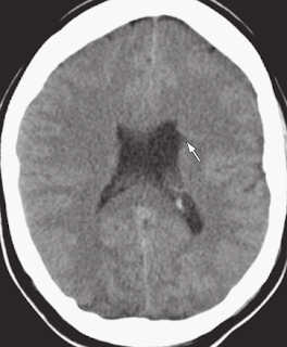
brain. A characteristic feature of such clefts is a slight outpouching or
“nipple” along the ependymal surface of the cleft. This is most often seen with
closed lip, or minimally open lip, schizencephaly. The gray matter lining the
cleft is always abnormal, and frequently demonstrates a nodular gyral pattern.
Less Common
Radiologic Manifestations
A large percentage of patients with schizophrenia present with
an absent septum pellucidum. Almost half of such patients exhibit optic nerve
hypoplasia. The neuroradiologist should therefore evaluate for septo-optic
dysplasia in patients with schizencephaly.Mild hypoplasia of the corpus
callosum is commonly seen.
Primary Differential
Diagnosis
1-Porencephalic Cyst
Discussion of
Differential Diagnosis
Porencephalic Cyst:
Cerebrospinal fluid-filled cyst lined by gliotic white matter and not gray
matter.
The patient is a 16-year-old boy with seizures.
Axial CT scan demonstrates a deep gray matter–lined (arrowheads) cleft extending from the pia of the overlying cerebral cortex to the ependymal lining of the ventricle.
At a slightly caudal level, axial CT scan demonstrates a slight outpouching or “nipple” (arrow) along the ependymal surface of the cleft. Diagnosis: Closed-lip schizencephaly.
Sponsored Links
|
Labels: Brain, Congenital Anomalies


